Anatomy Of Chest : Chest And Abdominal Anatomy Of A Child 2 Years Old Medical Stock Images Company. The thorax has two major openings: Computed tomography (ct) of the chest can detect pathology that may not show up on a conventional chest radiograph(1). Book of chest anatomy is a passive item. This tutorial is designed to help you understand the normal anatomy of the chest as seen on ct images in three planes: Anatomy of the thoracic wall.
Shop devices, apparel, books, music & more. 1 effects 2 notes 3 trivia 4 see also improves the contents of broken chests. (1) the pectoralis major, and (2) the pectoralis minor. Computed tomography (ct) of the chest can detect pathology that may not show up on a conventional chest radiograph(1). It provides a protective framework for…
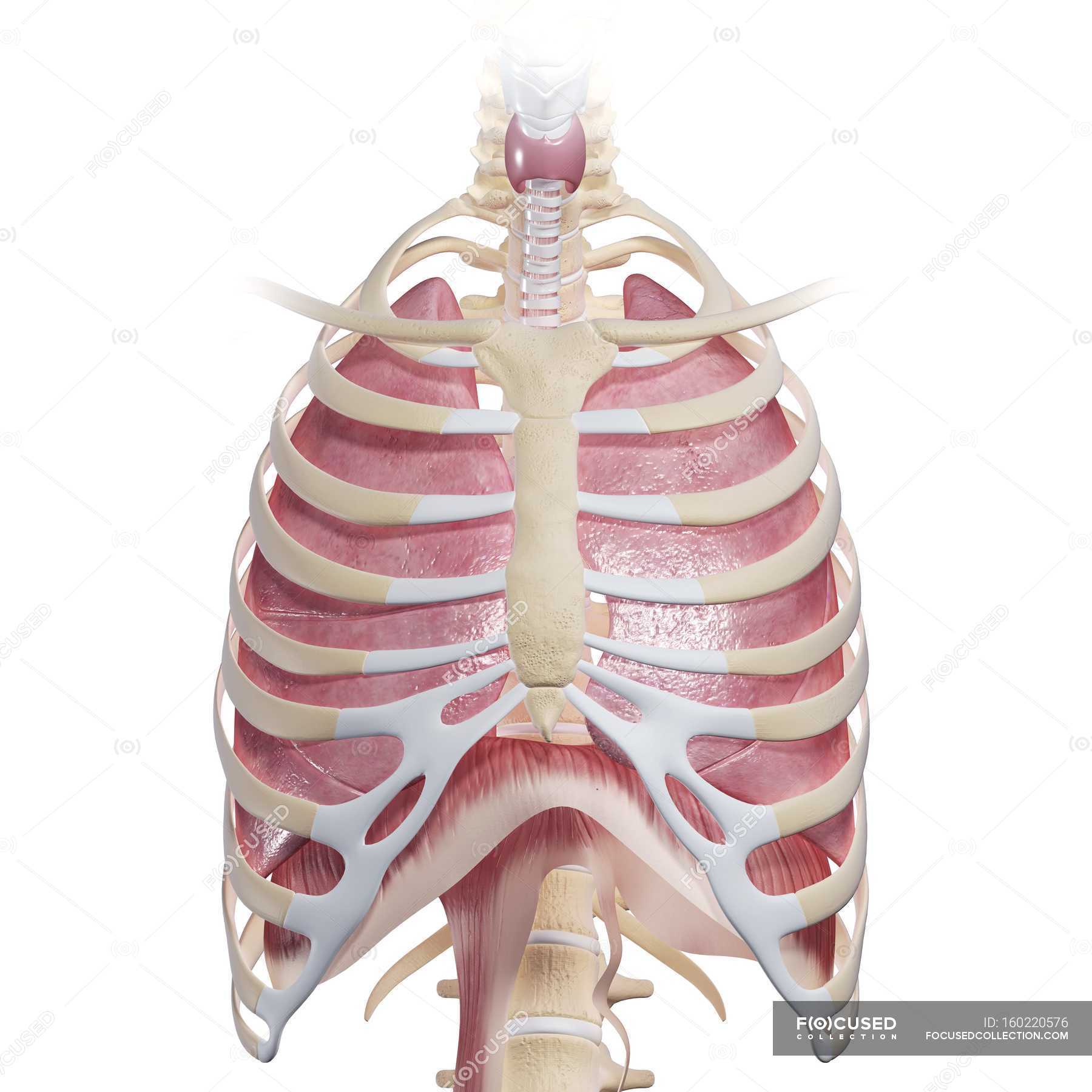
Human Chest Anatomy Close Up Anatomical Reference Stock Photo 160220576 from st.focusedcollection.com This atlas is a comprehensive and affordable learning tool for medical students and residents and especially for radiologists and pneumologists. Anatomy of the thorax, heart, abdomen and pelvis recommended text gray's anatomy for students. This page provides an overview of the chest muscle group. Search for human anatomy chest diagram. In particular, the right side of the chest is home to several structures including the right side of the heart, the three lobes of the right lung, the ascending aorta, the pulmonary blood vessels,. Free shipping on qualified orders. 1 effects 2 notes 3 trivia 4 see also improves the contents of broken chests. The first step in understanding thorax anatomy is to find out its boundaries.
A line is drawn from anterior surface of the body of 6th thoracic vertebrae passing through the apex of the heart up to anterior lower most part of diaphragm.
The heart is a muscle at the center of your circulatory system. Book of chest anatomy is a passive item. About the 6th week, the somites differentiate into the sclerotomes and the dermatomyotomes. The thoracic skeleton creates a protected space for the heart. The first step in understanding thorax anatomy is to find out its boundaries. How to view the anatomical labels. Search for human anatomy chest diagram. The epidermis is the outermost layer that provides a protective, waterproof seal over the body. In particular, the right side of the chest is home to several structures including the right side of the heart, the three lobes of the right lung, the ascending aorta, the pulmonary blood vessels,. The bony and soft tissue components of the chest wall combine to create an anatomic space, which houses some of the most vital structures in the human body. Thoracic cavity, also called chest cavity, the second largest hollow space of the body. It is enclosed by the ribs, the vertebral column, and the sternum, or breastbone, and is separated from the abdominal cavity (the body's largest hollow space) by a muscular and membranous partition, the diaphragm. Learn about each of these muscles, their locations, functional anatomy and exercises for them.
In particular, the right side of the chest is home to several structures including the right side of the heart, the three lobes of the right lung, the ascending aorta, the pulmonary blood vessels,. Anatomy of the thorax shari l. About the 6th week, the somites differentiate into the sclerotomes and the dermatomyotomes. A line is drawn from anterior surface of the body of 6th thoracic vertebrae passing through the apex of the heart up to anterior lower most part of diaphragm. The chest is the area of origin for many of the body's systems as it houses organs such as the heart, esophagus, trachea, lungs, and thoracic diaphragm.
Internal Anatomy Of Male Chest And Abdomen On Black Stock Photo Download Image Now Istock from media.istockphoto.com This section of the website will explain large and minute details of arterial anatomy of chest Swensen fund for innovation in teaching. Chest muscles anatomy (1) pectoralis major muscle. The circulatory system does most of its work. The chest wall is comprised of skin, fat, muscles, and the thoracic skeleton. A line is drawn from anterior surface of the body of 6th thoracic vertebrae passing through the apex of the heart up to anterior lower most part of diaphragm. This atlas is a comprehensive and affordable learning tool for medical students and residents and especially for radiologists and pneumologists. Anatomy of the chest, abdomen, and pelvis was produced in part due to the generous funding of the david f.
How to view the anatomical labels.
Studied the anatomy of the breast, its topography, innervation, vascularization and lymphatic drainage, and correlated the anatomical data with the classification of lymph node groups that is frequently utilized by mastologists. It provides protection to vital organs (eg, heart and major vessels, lungs, liver) and provides stability for movement. Search for human anatomy chest diagram. It provides a protective framework for… It is enclosed by the ribs, the vertebral column, and the sternum, or breastbone, and is separated from the abdominal cavity (the body's largest hollow space) by a muscular and membranous partition, the diaphragm. The chest anatomy includes the pectoralis major, pectoralis minor and the serratus anterior. An overview of the anatomy visible in a transverse computed axial tomographical image of the thorax (and part of the abdomen) performed with intravenous cont. Diseases of the chest and chest abnormalities make up a significant portion of a physician's daily practice. It spreads out like a fan and covers the rib cage like an armor plate. It provides access to ct images in the axial plane, allowing the user to learn and review the lung anatomy interactively. Heart your heart sits in the middle of your chest, to the left. Shop devices, apparel, books, music & more. 12 cm (5 in) in length, 8 cm (3.5 in) wide, and 6 cm (2.5 in) in thickness.
The major muscle in the chest is the pectoralis major. A line is drawn from anterior surface of the body of 6th thoracic vertebrae passing through the apex of the heart up to anterior lower most part of diaphragm. Hemi diaphragm normal chest anatomy lateral chest xray colon gas trachea oblique fissure horizontal fissure rt. 12 cm (5 in) in length, 8 cm (3.5 in) wide, and 6 cm (2.5 in) in thickness. Learn about each of these muscles, their locations, functional anatomy and exercises for them.
Chest And Abdominal Anatomy Of A Child 2 Years Old Medical Stock Images Company from cdn.shopify.com Book of chest anatomy is a passive item. Heart your heart sits in the middle of your chest, to the left. Radiology basics of chest ct anatomy with annotated coronal images and scrollable axial images to help medical students and junior doctors learning anatomy. Whatever you need, whatever you want, whatever you desire, we provide. In particular, the right side of the chest is home to several structures including the right side of the heart, the three lobes of the right lung, the ascending aorta, the pulmonary blood vessels,. Anatomy of the thorax, heart, abdomen and pelvis recommended text gray's anatomy for students. Learn about each of these muscles, their locations, functional anatomy and exercises for them. The thorax has two major openings:
1 effects 2 notes 3 trivia 4 see also improves the contents of broken chests.
Applied anatomy of the chest wall and mediastinum petros mirilas michael e. The skeleton of the thoracic wall is formed by the twelve thoracic vertebra posteriorly, It is enclosed by the ribs, the vertebral column, and the sternum, or breastbone, and is separated from the abdominal cavity (the body's largest hollow space) by a muscular and membranous partition, the diaphragm. It spreads out like a fan and covers the rib cage like an armor plate. The chest is made up primarily of two muscles: It provides protection to vital organs (eg, heart and major vessels, lungs, liver) and provides stability for movement. This atlas is a comprehensive and affordable learning tool for medical students and residents and especially for radiologists and pneumologists. Shop devices, apparel, books, music & more. In insects, crustaceans, and the extinct trilobites, the thorax is one of the three main divisions of the creature's body, each of which is in turn composed of multiple segments. The chest wall is comprised of skin, fat, muscles, and the thoracic skeleton. Anatomy of the thorax, heart, abdomen and pelvis recommended text gray's anatomy for students. About the 6th week, the somites differentiate into the sclerotomes and the dermatomyotomes. It provides a protective framework for…
Share :
Post a Comment
for "Anatomy Of Chest : Chest And Abdominal Anatomy Of A Child 2 Years Old Medical Stock Images Company"


Post a Comment for "Anatomy Of Chest : Chest And Abdominal Anatomy Of A Child 2 Years Old Medical Stock Images Company"